Which Interval/segment Observed via Ekg Sensor Can Be Used to Calculate the Heart Rate?
Man Physiology
Featured Human Physiology Categories
Featured Man Physiology Experiments
Introduction to Electrocardiography
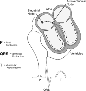
An electrocardiogram (ECG or EKG) is a graphical recording of the electrical events occurring within the center. In a healthy center at that place is a natural pacemaker in the right atrium (the sinoatrial node) which initiates an electrical sequence. This impulse then passes downwards natural conduction pathways betwixt the atria to the atrioventricular node and from in that location to both ventricles. The natural conduction pathways facilitate orderly spread of the impulse and coordinated contraction of first the atria and and then the ventricles. The electrical journey creates unique deflections in the EKG that tell a story about heart office and health. Fifty-fifty more data is obtained by looking at the story from different angles, which is accomplished by placing electrodes in various positions on the chest and extremities. A positive deflection in an EKG tracing represents electrical activity moving toward the active lead (the light-green pb in this experiment).
5 components of a single beat out are traditionally recognized and labeled P, Q, R, South, and T. The P moving ridge represents the starting time of the electrical journeying every bit the impulse spreads from the sinoatrial node downwardly from the atria through the atrioventricular node and to the ventricles. Ventricular activation is represented past the QRS circuitous. The T wave results from ventricular repolarization, which is a recovery of the ventricular musculus tissue to its resting land. By looking at several beats you tin can likewise calculate the rate for each component.
Doctors and other trained personnel tin expect at an EKG tracing and see show for disorders of the heart such equally abnormal slowing, speeding, irregular rhythms, injury to musculus tissue (angina), and death of muscle tissue (myocardial infarction). The length of an interval indicates whether an impulse is post-obit its normal pathway. A long interval reveals that an impulse has been slowed or has taken a longer route. A short interval reflects an impulse which followed a shorter route. If a complex is absent, the electrical impulse did non ascension usually, or was blocked at that part of the heart. Lack of normal depolarization of the atria leads to an absent P wave. An absent QRS complex after a normal P moving ridge indicates the electrical impulse was blocked earlier it reached the ventricles. Abnormally shaped complexes result from aberrant spread of the impulse through the muscle tissue, such as in myocardial infarction where the impulse cannot follow its normal pathway because of tissue death or injury. Electrical patterns may besides be changed by metabolic abnormalities and by various medicines.
In this experiment, you volition employ the EKG sensor to make a five second graphical recording of your center's electric activity, and so switch the red and green leads to simulate the change in electric activity that can occur with a myocardial infarction (heart attack). You volition identify the unlike components of the waveforms and use them to determine your heart rate. You lot volition also decide the direction of electrical activity for the QRS circuitous.
Read More »
Limb Position and Grip Strength
The importance of manus strength and function is axiomatic in all aspects of our daily living, from eating and maintaining personal hygiene to typing at the computer, performing brain surgery, or playing tennis or the piano. People suffering from arthritis or mitt injury apace capeesh the difficulty of performing even simple tasks with reduced grip strength.
Testing of hand grip forcefulness is used by orthopedic surgeons and concrete therapists to evaluate the extent of an injury and the progress of recovery. Grip force tin besides be used to diagnose neuromuscular problems such as stroke, herniated disks in the neck, carpal tunnel syndrome, and elbow tendonitis. Athletes are interested in grip strength because it relates to performance in many sports, such as tennis, golf, baseball game, football game, gymnastics, and rock climbing.
Compression strength is a style for occupational therapists to mensurate loss of fine-motor strength in the thumb, fingers, and forearm. It is useful for analyzing the extent of an injury and the event from surgery or therapy.
In Part I of this experiment, you will measure and compare grip strength in your correct and left hands. You volition also correlate grip strength with arm position, handedness, and height. In Part 2 you will analyze the pinch strength of each of your four fingers.
Read More »
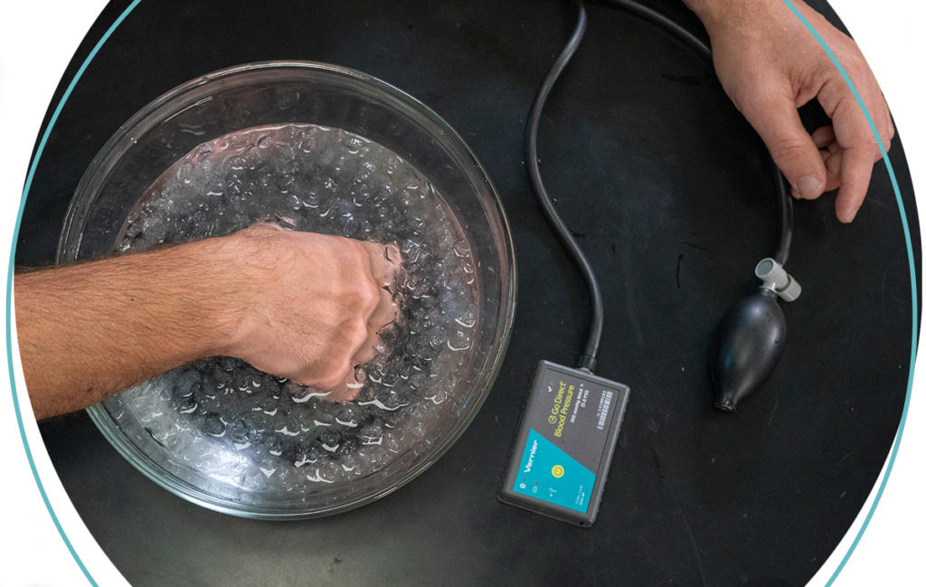
Effect of Exercise on Centre Rate
The adaptability of the heart can exist observed during do, when the metabolic activity of skeletal muscle tissue increases. The cardiovascular organisation, consisting of the heart and blood vessels, responds to exercise with an increase in middle rate and strength of contraction with each beat, resulting in a higher cardiac output (quantity of blood pumped through the heart per unit of time). Physically fit people can deliver a greater book of blood in a single heartbeat than unfit individuals and can sustain a greater work level before reaching a maximum middle charge per unit. Being more physically fit too leads to a more rapid recovery of resting heart charge per unit.
In this experiment, you volition observe how the middle responds to the increased metabolic demand of muscles during exercise.
Read More than »
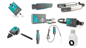
Featured Physiology Products
Source: https://www.vernier.com/human-physiology/
0 Response to "Which Interval/segment Observed via Ekg Sensor Can Be Used to Calculate the Heart Rate?"
Post a Comment